

Research on Medical Imaging
Segmentation of the IAN Canal in CBCT Volumes

In maxillofacial imagery, recent works have been focused on the detection of the Inferior Alveolar Nerve (IAN), since its position is of great relevance for avoiding severe injuries during surgery operations such as third molar extraction or implant installation.
Our efforts on this research area have been focused on:
- Creating novel tools for analyzing and labeling the alveolar nerve from Cone Beam Computed Tomography (CBCT) 3D volumes;
- Releasing valid datasets for deep learning purposes;
- Designing 3D neural networks for the automatic segmentation of the IAN;
Skin Lesion Analysis
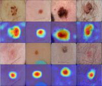
Skin lesion analysis has been one of the research fields of our labs for several years.
In particular, we made use of Convolutional Neural Networks (CNNs) to perform semantic segmentation and lesion classification on dermoscopic images. Moreover, we explored the usage of Generative Adversarial Networks (GANs) in the data augmentation process, and developed a tool for content-based dermoscopic image retrieval.
Beside writing numerous published papers we participated to the International Skin Imaging Collaboration (ISIC) challenge, achieving the third best result in 2019 in collaboration with the Universidad Politecnica de Valencia (UPV).
Deep Renal Biopsy Immunofluorescence Image Analysis
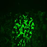
Immunohistopathology is an essential technique in the diagnostic workflow of a kidney biopsy, we wanted to evaluate the role of a Convolutional Neural Network (CNN) as a support tool for kidney immunofluorescence reporting. High magnification (400X) immunofluorescence images of kidney biopsies performed from the year 2001 to 2018 were collected. The report, adopted at the Division of Nephrology of the AOU Policlinico di Modena, describes the specimen in terms of 'Appearance', 'Distribution', 'Location' and 'Intensity' of the glomerular deposits identified with fluorescent antibodies against IgG, IgA, IgM immunoglobulins, C1q and C3 complement fractions, Fibrinogen, kappa and lambda light chains. The report was used as ground truth for the training of the CNNs. 12259 immunofluorescence images of 2542 subjects undergoing kidney biopsy were collected, the test set analysis showed accuracy values between 0.79 ('Irregular Capillary wall' feature) and 0.94 ('Fine Granular' feature). The agreement test of the results obtained by the Convolutional Neural Networks with respect to the ground truth showed similar values to three pathologists of our center. Convolutional Neural Networks were 117.3 times faster than human evaluators in analyzing 180 image tests. A web platform, where it is possible to upload digitized images of immunofluorescence specimens, is available to evaluate the potential of our approach.
3D Reconstruction of Skin Lesions for Tumor Diagnosis in OCT imaging

Optical Coherence Tomography (OCT) allows to capture the internal structure of skin lesions. In this project we develop an image analysis pipeline to obtain 3D models from OCT scans. Both the external surface and the internal vascular network are reconstructed to give to the clinicians new qualitative tools for tumor diagnosis.
This project is developed in conjunction with the dermathologists of Policlinico di Modena under the direction of Prof. Giovanni Pellacani.
This research is funded by the European Project ADVANCE: Automatic Detection of VAscular Networks for Cancer Evaluation.